


The brain is the organ that runs everything: movement, sensation, emotion, memory, language, sleep, and every decision you make without noticing. It consumes roughly 20% of your body’s energy despite being about 2% of your body weight. Many mechanisms that work differently in neurodivergent brains — from how you pay attention and how you process emotion to how you sleep — can often be traced back to differences in how this organ is structured, how it communicates, and what it needs to function well.
How to use this guide
Most of us last thought about how our brains work somewhere around Year 9 biology. We learned that mitochondria are the powerhouse of the cell, that neurons fire electrical signals, and that the brain has different lobes that do different things. Then most of us filed it all under “exam material” and moved on.
If you’re here, something has probably changed since then. Maybe you’ve recently learned you have a neurodivergent brain, or you’re starting to suspect you might. Either way, you’re looking at decades of your own experience through a new lens, and suddenly the biology feels personally relevant in a way it never did in a classroom.
Please note: This page is a refresher, not a comprehensive textbook chapter. We’ve gone through what’s known about brain biology and pulled out the parts that are the most relevant when you’re trying to understand neurodivergent traits: the structures, chemistry, networks, and the nutrients.
Table of Contents[Hide][Show]
How the brain communicates
Your nervous system is built from neurons, which are cells that send and receive electrical and chemical signals. They exist everywhere: the brain, spinal cord, and body. The neurons in your fingertips that detect heat are the same type of cell as the neurons in your prefrontal cortex that help you plan your week. Your brain contains roughly 86 billion of them. Your gut contains around 500 million more, using many of the same chemical messengers, which is why the gut is sometimes called the second brain.
A single neuron has a cell body (where its DNA and core machinery sit) and an axon — a long, thin extension that carries electrical signals away from the cell body toward the next cell. Some axons are fractions of a millimetre long. Others, like the sensory neurons running from your spinal cord to your feet, are over a metre. When thousands of axons are bundled together into a single cable, that cable is called a nerve. The vagus nerve, for example, is a bundle of roughly 100,000 axon fibres connecting the brainstem to the gut and other organs.
How neurons pass on information
Neurons, contrary to simplified descriptions, don’t communicate using either electricity or chemistry. They use both, everywhere in the body.
An electrical signal (called an action potential) travels down the length of the axon — that’s how the message moves within a single cell. When it reaches the end, the neuron releases chemicals called neurotransmitters across a tiny gap (the synapse) to the next cell. The receiving cell picks up those chemicals and decides whether to fire its own electrical signal. This mechanism — electrical along the axon, chemical across the gap — is how all neurons communicate, whether they’re in the brain, the spinal cord, the gut, or the tips of your fingers.
Neurons and neurotransmitters
Neurons are largely specialised.
- A dopaminergic neuron produces dopamine.
- A serotonergic neuron produces serotonin.
- A GABAergic neuron produces GABA.
This is why you’ll hear people talk about dopamine pathways and serotonin systems — they’re physically distinct populations of cells running through specific routes in the brain. Some neurons can release more than one neurotransmitter, but the general principle is specialisation. A neuron’s identity is partly defined by which chemical messenger it makes.
Different neurotransmitters do different jobs.
- Dopamine is involved in motivation, reward, and the ability to sustain attention on something that isn’t immediately interesting.
- Serotonin plays a role in mood, anxiety, sleep, and has a huge presence in our guts.
- Norepinephrine (also called noradrenaline) regulates alertness and focus, and is the target of some ADHD medications.
- GABA and glutamate work as a pair: GABA slows signalling down, glutamate speeds it up. The balance between them shapes how excitable or how calm your nervous system is at any given moment.19
These aren’t the only chemical messengers involved. Endorphins are the body’s own pain relief system, which is part of why intense exercise can feel regulating. Acetylcholine supports memory and learning. The brain also responds to hormones like cortisol (involved in stress) and oxytocin (involved in bonding and social connection). The boundary between neurotransmitters and hormones is not clear-cut, and both systems shape how you feel and function day to day.
How a brain signal travels
It’s easy to imagine neurotransmitters being passed along a chain like a baton in a relay race, but that’s not how it works. A neuron fires an electrical signal down its own length. When that signal reaches the end, the neuron releases its neurotransmitter into the synaptic gap. That neurotransmitter binds to receptors on the next neuron. If the signal is strong enough, the receiving neuron fires its own electrical signal, and at the end of that neuron, it releases its own neurotransmitter, which might be a completely different chemical. The original molecules don’t travel onward. They’re either reabsorbed by the neuron that released them — this is called reuptake, and it’s the mechanism that SSRIs and some ADHD medications interfere with — or they’re broken down by enzymes in the gap.
Each link in the chain is a fresh production event. Synthesis — the creation of a neurotransmitter molecule from scratch — happens every time a neuron needs to signal. Dopamine is synthesised from the amino acid tyrosine, in a process that requires iron at the critical first step. Serotonin is synthesised from tryptophan, another amino acid that comes from food. GABA synthesis requires vitamin B6. Some of the released molecules get reabsorbed and reused, which reduces the manufacturing burden, but the system is more similar to a production line, not a storage cupboard.
This is why what your brain needs to function is mechanically central and crucial to understand, and it’s not as simple as taking some supplement off eBay as a life hack. For example, if a neuron can’t access enough iron, it can’t produce dopamine at the rate it needs to, even if the neuron itself is perfectly healthy. The brain isn’t just using these chemicals. It’s constantly building them, and it needs a steady supply of the right raw materials to keep doing so.
Mini vocabulary
Amino acid — a small molecule that the body uses as a building block. Some come from food (like tyrosine from protein), and some the body makes on its own. Neurotransmitters are built from amino acids.
Axon — the long, thin extension of a neuron that carries electrical signals away from the cell body toward the next cell. Axons can be fractions of a millimetre or over a metre long. When thousands of axons are bundled together, the bundle is called a nerve.
Enzyme — a protein that makes a specific chemical reaction happen. Enzymes don’t get used up in the process — they do the same job over and over. Tyrosine hydroxylase is the enzyme that converts tyrosine into the first step of dopamine.
Cofactor — a helper molecule that an enzyme needs in order to work. Iron is a cofactor for tyrosine hydroxylase. Without the cofactor, the enzyme is present but can’t do its job.
Precursor — a substance that gets converted into something else. Tyrosine is a precursor of dopamine. Tryptophan is a precursor of serotonin. The precursor isn’t the finished product — it’s the raw material one step before.
How this plays out in the neurodivergent brain
In neurodivergent brains, these systems are tuned differently.
ADHD is closely associated with differences in how dopamine and norepinephrine work in the brain’s reward, motivation, and attention circuits.12
Autism involves differences in the balance between GABA and glutamate, which can show up as sensory sensitivity, intense focus, or the experience of a nervous system that doesn’t have a comfortable middle gear.19
These aren’t broken systems; they are just running on different settings.
The brain has dozens of identifiable regions, but the main two that will come up when you’re reading about ADHD and autism are the prefrontal cortex and the amygdala. They’re behind many of the traits and experiences that bring people to a diagnosis in the first place.
The prefrontal cortex and executive function
The prefrontal cortex sits at the front of the brain and is involved in planning, decision-making, impulse control, working memory, and the ability to regulate your own behaviour and emotions. These capacities are often grouped under the term executive function.
The prefrontal cortex is one of the last brain regions to fully mature — synapse formation, pruning, and neurotransmitter system refinement continue into our mid-twenties.45
This extended timeline means the prefrontal cortex is especially sensitive to differences in neurodevelopment.
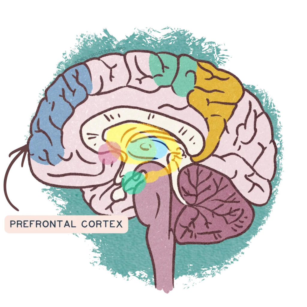
In ADHD, prefrontal volume and activation tend to be reduced, particularly in regions involved in cognitive control and emotional regulation.79 In autism, early childhood often shows larger-than-typical frontal lobe volume, which then follows a distinct trajectory over time.6 When ADHD and autism co-occur, the pattern is not simply a combination of the two — it’s its own distinct profile, which is part of why AuDHD can feel like a different experience from either condition alone.78
The prefrontal cortex is also where dopamine and norepinephrine do much of their work on attention and self-regulation. And when it comes to neurodivergent brains, even though the structure is there, the neurotransmitters it relies on are tuned differently.
The amygdala and emotional regulation
The amygdala is a small, almond-shaped structure deep in the brain, and it’s often described as the brain’s threat detector. It processes fear, anxiety, and emotional reactions, and it plays a role in how quickly and how intensely you respond to emotional situations.
In pop neuroscience, the amygdala gets treated as though it’s acting alone — “your amygdala got hijacked“. As is often the case, the real mechanics are more complex.
Emotional regulation is about the conversation between the amygdala and the prefrontal cortex. First, the amygdala flags something as emotionally relevant. The prefrontal cortex then evaluates it, contextualises it, and decides what to do about it. When that conversation works smoothly, you can feel an emotional reaction and modulate your response. When the connection between the two is different — as it is in both ADHD and autism, though in different ways — the emotional reaction arrives faster or more intensely than the prefrontal cortex can manage.
Emotional regulation in ADHD & Autism
In adult ADHD, emotional dysregulation affects roughly 30–70% of adults,1011, and the research consistently points to altered connectivity between the amygdala and medial prefrontal cortex rather than the amygdala “overreacting” in isolation.1218
Adults with ADHD, especially if they have no support systems established, tend to use less cognitive reappraisal (reframing what happened) and rely more on suppression (pushing the feeling down), which is a less effective long-term strategy.1314

In autism, the picture is more mixed. A large meta-analysis found consistent underactivation of the amygdala during negative emotional processing,15 while structural studies show larger amygdala volume and atypical connectivity with social processing regions.16 One recent study in autistic adults found no difference in amygdala activation at all during basic emotional face processing17 — which challenges the simple “overactive amygdala” narrative and suggests the differences are in how the amygdala connects to the rest of the brain, not in how “reactive” it is.
The traits you may experience, like difficulty planning things, emotional overwhelm, meltdowns, and the sense that your reactions are too fast for your own self-regulation to catch, all have identifiable biology behind them. Knowing where they come from doesn’t fix them, but it can change the story you tell yourself about why they happen.
The default mode network
Not all brain activity is about responding to the outside world.
When you’re not focused on a specific task — for example, when you’re daydreaming, replaying a conversation, thinking about the future, or just staring out of a window — a group of brain regions becomes more active rather than less. This group is called the default mode network, and it was only identified in the early 2000s. It includes the medial prefrontal cortex, the posterior cingulate cortex, and parts of the parietal and temporal lobes.

The default mode network is involved in self-referential thinking (thoughts about yourself, your past, your plans), social cognition (imagining what someone else might be thinking or feeling), and the kind of free-form mental wandering that people sometimes call “being in your own head.” It’s also meant to quiet down when you switch to a focused, goal-directed task — handing over to what researchers call the task-positive network. In practice, your brain is constantly shifting between these two modes depending on what’s being asked of it.
What the DMN is doing differently in neurodivergent brains
In both ADHD and autism, the default mode network works differently, though not in the same way.2021
In ADHD, the default mode network often stays active when it should be quieting down. Instead of handing over cleanly to the task-positive network, the two compete.2025 This is one of the reasons people with ADHD describe their minds as “always on” — the network responsible for internal chatter doesn’t step back when external focus is needed. If you’ve ever been reading a paragraph and realised three sentences in that you’ve been thinking about something else entirely, that’s what impaired DMN suppression feels like from the inside.
In autism, the pattern is more mixed. Some studies find reduced connectivity within the default mode network itself, particularly between the regions involved in social cognition.2223 Others find increased connectivity between the default mode network and other networks, including the salience network, which decides what deserves attention.2125 The overall picture is that the DMN in autistic brains is organised differently, rather than simply overactive or underactive, and these differences relate to social processing and to the way attention is allocated between internal and external experience.
When ADHD and autism co-occur, the default mode network shows more extreme alterations than either condition on its own24 — further evidence that AuDHD is its own distinct neurological profile rather than one condition layered on top of another.
Default mode network differences alone don’t cleanly separate ADHD from autism.202125 Both conditions show atypical DMN patterns, and there’s overlap. The main thing you need to understand is this: the brain has a network that governs what your mind does when nothing external is demanding its attention, and in neurodivergent brains, that network operates on different rules.
What your brain needs
Your brain is a manufacturing operation. Every signal it sends requires raw materials — amino acids, minerals, vitamins — to build neurotransmitters. When those materials are in short supply, the production line slows down. The brain doesn’t stop working, but it works less efficiently, and the effects show up in exactly the places neurodivergent people already struggle with: attention, motivation, sleep, emotional regulation, and processing speed.
Research consistently finds that people with ADHD, autism, and other neurodivergent conditions are more likely to have lower levels of several key nutrients, including iron, magnesium, zinc, vitamin D, B-vitamins, and omega-3 fatty acids.37 This doesn’t mean deficiency causes neurodivergence. It means neurodivergent brains may need more attention in these areas, and that unaddressed deficiencies can make existing traits harder to manage.
Iron and dopamine
Iron is the nutrient with the clearest mechanistic connection to neurodivergent experience. It’s a required cofactor for tyrosine hydroxylase, the enzyme that converts the amino acid tyrosine into the precursor of dopamine. Without enough iron, this enzyme can’t do its job at full capacity, and dopamine production slows down. This doesn’t just reduce how much dopamine is available — it affects the entire reward and motivation circuit that dopamine supports.
Brain imaging studies have consistently found reduced iron concentrations in the basal ganglia — which are the brain regions most involved in dopaminergic signalling — of children with ADHD. A 2024 systematic review of seven neuroimaging studies found this to be a consistent finding across different MRI techniques and different populations.26 Lower brain iron correlated with more severe ADHD symptoms, particularly inattention and difficulties with inhibitory control.2640
If you have been told your blood work is normal, but you still feel odd, this is probably going to vindicate your instincts: brain iron and blood iron are not the same measurement, and they don’t always agree. Serum ferritin — the standard blood test used to assess iron stores — often does not correlate well with MRI-estimated brain iron in ADHD.262728 You can have a ferritin level that falls within the laboratory reference range and still have meaningfully low iron in the brain regions that need it for dopamine production.
This discrepancy is made worse by the fact that ferritin is what’s called an acute-phase reactant. When your body is fighting inflammation — even low-grade, chronic inflammation — ferritin levels go up independently of your actual iron stores.31 Research estimates this can raise ferritin by 30–50%, leading to a 10–30% underestimation of iron deficiency at the population level.2930 This means the standard test can look reassuring even when the situation is far from it.
Iron, periods and ADHD
Anyone who menstruates loses iron through menstrual blood loss every month, and this creates a compounding problem for people with ADHD specifically. Research shows that PMDD, heavy menstrual bleeding (menorrhagia), dysmenorrhea, and PCOS all occur at higher rates in women with ADHD than in the general population.42434446 One large survey study found the odds of PMDD were roughly 6,5 times higher in women with ADHD.43 In a community sample, women with symptoms suggestive of ADHD were more likely to report heavy menstrual bleeding and symptoms of iron deficiency than those without (39% compared with 26%).44 The population most likely to have dopamine-related attention difficulties is also the population most likely to be losing more iron than average.
Why ADHD traits seem worse with the menstrual cycle
To make it absolutely clear: this doesn’t mean iron deficiency is being mistaken for ADHD and causes overdiagnosis of ADHD. The ADHD exists, and it’s there all month. But iron loss during menstruation can make existing symptoms measurably worse for part of the cycle. Multiple studies report that women with ADHD experience worsening attention, mood, and executive function in the late luteal and premenstrual phase, and many describe uncertainty about whether their medication is still working during this window.424745 If you notice your stimulant medication seems to stop working for a week or two each month, then starts working again, this in itself can be a clue. If medication resistance were caused by ADHD alone, it would be consistent. When it’s cyclical, something else is contributing — and iron depletion through menstrual blood loss is one of the most plausible and most overlooked mechanisms.
A blinded, placebo-controlled trial in women aged 18–35 found that iron status directly affected cognitive performance. Iron-sufficient women performed better on cognitive tasks and completed them faster than women with iron deficiency. After 16 weeks of supplementation, improvement in ferritin was associated with a 5–7-fold improvement in cognitive accuracy, while improvement in haemoglobin was associated with faster processing speed.32 Even mild low ferritin in non-anaemic participants — people who wouldn’t be flagged as deficient by standard screening — was associated with needing more time to complete cognitive tasks.33
For late-identified neurodivergent women, you might be familiar with this storyline: your ADHD symptoms fluctuate with the menstrual cycle, and your heavier-than-average periods were never connected to ADHD. Your blood work comes back “normal” because the ferritin reference range doesn’t account for neurobiological function. You are left with a growing sense that something still doesn’t add up. The ADHD diagnosis is still valid, but a possible iron insufficiency that is making it worse has been overlooked.
Iron, restless legs, and sleep
The iron-dopamine connection also explains a co-occurring condition. Roughly 20–35% of adults with ADHD meet criteria for restless legs syndrome — the urge to move your legs that worsens at rest and at night.35
A Mendelian randomisation study found evidence that ADHD is an independent causal risk factor for developing RLS, but not the reverse.34 The shared reason appears to be brain iron deficiency: low iron in the substantia nigra and thalamus disrupts dopamine regulation in a circadian pattern, producing a hyperdopaminergic state during the day that gives way to relative dopamine underactivity at night, when RLS symptoms peak.36
If you’ve experienced the combination of restless legs, fragmented sleep, morning exhaustion, and daytime attention problems — and been told these are separate issues — the research suggests they may be connected through the same iron-dopamine pathway that is affecting your other traits.
Other nutrients important for neurodivergent brains
Iron’s role in ADHD has the deepest evidence base, but it isn’t the only nutrient that shows up repeatedly in the research.
Magnesium levels are frequently lower in children and adults with ADHD.41 Magnesium is involved in GABA function, stress regulation, and sleep. Supplementation studies show modest improvements in hyperactivity, attention, and emotional difficulties, particularly when deficiency is present — but the evidence isn’t strong enough to recommend it as a standalone treatment.
Vitamin D deficiency is more prevalent among autistic children than non-autistic peers, with some studies reporting rates above 50%.39 Several cohort studies link low maternal or neonatal vitamin D to higher autism risk, though cause and effect aren’t established. If lower vitamin D is more common in autism, and autism is hereditary, the autistic mother has lower vitamin D levels, and so will the child that inherits her autism trait bundle. The safest interpretation is that low vitamin D is a common co-occurrence that’s good to know about and check occasionally.
Omega-3 fatty acids, zinc, and B vitamins all appear in the research as nutrients that tend to be lower in neurodivergent populations.37 Supplementation trials show mixed results, and the evidence doesn’t support blanket recommendations. What the research does support is that structured nutritional assessment — a proper look at what someone is actually eating and absorbing, not just a routine blood panel — is worth doing when someone is neurodivergent, because the baseline risk of insufficiency is higher.
Most of the detailed nutritional research is in children. The evidence in autistic adults specifically is sparse — one recent review found only 43 relevant studies across all databases.38 Autistic adults tend toward sensory-driven food choices that can lead to nutrient-poor diets and higher rates of deficiency, but true absorption differences haven’t been well measured.38 This is a gap in the research rather than an absence of a problem.
What crosses the blood-brain barrier
Between your bloodstream and your brain tissue sits a tightly controlled gateway called the blood-brain barrier. It’s a layer of specialised cells lining the brain’s blood vessels, sealed together with structures called tight junctions, and its job is to be selective: letting in what the brain needs (oxygen, glucose, amino acids, certain nutrients) and keeping out what could harm it (toxins, pathogens, most of what’s circulating in your blood at any given moment).
The BBB determines which substances actually reach the organ. You can swallow something, digest it, absorb it into your bloodstream, and still have it never reach your brain — or reach it only partially, or reach it in ways that depend on your individual biology. The blood-brain barrier is the reason why what you eat, drink, and take as medication affects your brain differently from how it affects the rest of your body.
How medications get through
Most psychiatric medications, including stimulants prescribed for ADHD, are designed to be small and lipid-soluble enough to cross the blood-brain barrier through passive diffusion. Once across, stimulants like methylphenidate block the reuptake of dopamine in the ventral striatum and prefrontal cortex, increasing the amount of dopamine available in the synapse. Research using PET imaging in adults with ADHD has shown that the degree of dopamine increase in the ventral striatum — the brain’s reward and motivation region — predicted how much symptoms of inattention improved over 12 months of treatment.3
But getting a drug across the barrier is more complicated than that. The blood-brain barrier has active efflux pumps — proteins that actively push certain substances back out of the brain after they’ve crossed in.4951 Many antipsychotics, for example, are substrates of these pumps, which can lower the amount of drug that actually reaches brain tissue and contribute to variable treatment responses. Genetic variants in the genes coding for these pumps can mean that two people taking the same medication at the same dose end up with meaningfully different amounts reaching their brains.51 Sex hormones can also modify how these transporters work, adding another layer of individual variation.50
Everyday substances and your brain chemistry
Medications aren’t the only things crossing the blood-brain barrier. Several substances people consume daily interact with brain chemistry in ways that are directly relevant to the neurodivergent experience. A chemical is a chemical is a chemical, so whether it is in a pill or in a cup, it can have an effect on your brain.
For how everyday substances like caffeine, alcohol, and nicotine interact with the same barrier and the same brain chemistry, see our entry on self-medication.
BBB differences in neurodivergent conditions
Research on the blood-brain barrier in neurodivergent conditions is still in its early stages, but what exists is suggestive. The strongest evidence comes from autism, where postmortem brain tissue shows altered expression of tight junction proteins (the structures that seal the barrier) in both the cortex and cerebellum, coupled with increased markers of neuroinflammation.48 In the gut, the same study found that 75% of autistic tissue samples showed reduced barrier-forming components in the intestinal wall, while 66% showed increased pore-forming components — suggesting that both the gut barrier and the brain barrier may be more permeable than typical.48 For ADHD, preliminary biomarker studies hint at blood-brain barrier changes, but strong evidence is still lacking.49 This is an area where the research is moving but hasn’t arrived yet.
The gut-brain axis & the vagus nerve
Your gastrointestinal tract has its own nervous system, the enteric nervous system, containing hundreds of millions of neurons that use many of the same neurotransmitters as the brain. The main communication line between the two is the vagus nerve, which carries roughly 80% of its traffic from the gut to the brain rather than the other way around.63 Through its efferent fibres, the vagus nerve operates the cholinergic anti-inflammatory pathway, which dampens inflammation and helps regulate how tightly sealed the gut wall is.63 Chronic stress suppresses this pathway,6367 affecting digestion, immune function, and mood simultaneously.
Approximately 90% of the body’s serotonin is produced in the gut by specialised cells in the intestinal lining.62 Gut bacteria influence how much serotonin these cells produce, and the signals travel up the vagus nerve to brain regions involved in mood and emotional regulation.62
Gastrointestinal symptoms are reported in 25–70% of autistic individuals.65 Sensory-driven food selectivity can lead to restricted diets that alter the gut microbiota, which worsens GI symptoms, which further restricts food choices — a feedback loop in which autistic traits, eating, the microbiome, and gut function all shape each other.38
The vagus nerve’s role in this system has attracted a lot of oversimplification, particularly around cold showers and quick-fix “vagal toning.” Clinical vagus nerve stimulation requires a surgically implanted device delivering calibrated electrical pulses and is approved for treatment-resistant epilepsy and depression.67 Transcutaneous vagus nerve stimulation (tVNS) is being researched, but the evidence base is still developing.68 Slow diaphragmatic breathing does increase vagal tone temporarily and can help with acute stress regulation69, but the idea that a brief cold shower can meaningfully reset a system this complex doesn’t follow from the biology.70
For a full exploration of the gut-brain axis, including the gut microbiome, intestinal permeability, and the evidence on probiotics, see the dedicated gut-brain axis glossary entry.
Neuroplasticity
If everything above sounds fixed — structures, pathways, networks, barriers — let’s end this entry on the fact that, luckily, it isn’t. Your brain changes throughout your life. It forms new synaptic connections, strengthens the ones that get used, and through a process called synaptic pruning, removes the ones that don’t. This capacity is called neuroplasticity, and it doesn’t stop after childhood. Yes, it slows down, but it continues into adulthood and old age.
Neuroplasticity is the reason you can learn a new skill at 45, adjust to a major life change at 60, or develop coping strategies after a late diagnosis that change how you function day to day. Every time you practise something — a new habit, a new way of responding to a situation, a new way of organising your environment — you’re shaping the physical structure of your brain. The synaptic connections supporting that behaviour get stronger, and the ones you stop using get weaker.
Neuroplasticity in neurodivergent brains
In neurodivergent brains, plasticity works differently, but that doesn’t mean it works less. The research suggests the opposite in some cases: autistic brains can show hyperplasticity — a heightened capacity for synaptic change in certain domains.71 This may contribute to the intense skill acquisition and deep knowledge development that many autistic people experience in areas of strong interest, and it may also be part of why sensory environments that change unpredictably can feel overwhelming. A system that responds more intensely to input will respond more intensely to all input, including input you didn’t choose.
In ADHD, plasticity-related research has focused on how the brain responds to behavioural interventions, cognitive training, and medication. Stimulant medication appears to influence brain structure over time — the Morandini review discussed earlier found that psychostimulant use was associated with normalised brain iron levels in basal ganglia regions, and longer treatment duration correlated with greater normalisation.26 This suggests medication isn’t just managing symptoms in the moment. It may be supporting the underlying conditions the brain needs to function and adapt.
What helps with neuroplasticity
Movement
The single most evidence-based thing you can do to support neuroplasticity is aerobic exercise. Sustained aerobic activity — 30 minutes or more, three or more times a week.
Aerobic means anything that raises your heart rate and keeps it up for a sustained period. Walking at a pace that makes you slightly breathless counts. So does dancing, cycling, swimming, gardening that involves real digging or raking, or chasing your kids around a park. You don’t need a gym membership or a running habit. All you need is movement that gets your heart working harder than it does when you’re sitting down, maintained long enough for the body to respond to it.
Aerobic exercise increases production of brain-derived neurotrophic factor (BDNF), a protein that supports the survival of existing neurons, encourages the growth of new synaptic connections, and is involved in long-term memory formation.7475 The effects have been demonstrated in adults with neurological conditions,73 in healthy ageing populations, and in both ADHD and autism research specifically.
Nutriton
BDNF production is also influenced by omega-3 fatty acid status,78 and neurotransmitter synthesis — dependent on iron, B-vitamins, and amino acids — provides the chemical foundation that plasticity operates on top of. A brain that’s short on raw materials for neurotransmitter production has less to work with when it’s trying to form and strengthen new connections.
Sleep
During sleep — particularly deep slow-wave sleep — the brain consolidates learning, clears metabolic waste products, and carries out synaptic maintenance.7677 The sleep difficulties common in both ADHD and autism (delayed sleep phase, fragmented sleep, difficulty with sleep onset) aren’t just quality-of-life problems. They directly affect the brain’s capacity to adapt and reorganise. The iron-dopamine-sleep connection described earlier on this page is relevant here too: disrupted sleep doesn’t just make you tired the next day. It reduces the window in which neuroplasticity can do its work.
What neuroplasticity means for late-identified adults
For people who receive a neurodivergent diagnosis in their thirties, forties, or later, one of the most common questions is: Is it too late? The neuroscience is clear that it isn’t. The brain you have now is not the brain you’re stuck with. It is capable of forming new connections, strengthening useful patterns, and adapting to new understanding of itself.
Neuroplasticity doesn’t mean you can think your way out of a neurological difference, and it doesn’t mean the right mindset can substitute for medication, support, or accommodations. What it does mean is that the strategies you build, the environments you choose, the sleep you protect, and the understanding you develop about how your own brain works — all of these produce physical changes in your brain’s structure over time. Late diagnosis isn’t a closed door. Try to think of it as a different starting point, where you still have options for different trajectories.
« Back to the index





 “Can supplements help with ADHD?”